Bone Cross Section View - Human Skull Mid Sagittal Cross Section Side View On White Background Stock Photo Picture And Royalty Free Image Image 120521964 / They are obtained by taking imaginary slices perpendicular to the main axis of organs, vessels, nerves, bones, soft tissue, or even the entire human body.
Bone Cross Section View - Human Skull Mid Sagittal Cross Section Side View On White Background Stock Photo Picture And Royalty Free Image Image 120521964 / They are obtained by taking imaginary slices perpendicular to the main axis of organs, vessels, nerves, bones, soft tissue, or even the entire human body.. Start studying bone cross sections. 320 × 160 pixels | 640 × 320 pixels | 1,024 × 512 pixels | 1,280 × 640 pixels | 1,000 × 500 pixels. For example, if there was a hollow cavity, or internal features, or. Do not color the articular cartilage. Size of this png preview of this svg file:
We can see there are two layers of compact bone here. Two pulp canals are visible in the mesiobuccal root of the first molar. Color the bone tissue gold. Different types of bones from passassignment2014.weebly.com there are trabeculae in spongy bone. Section of bone marrow affected by myeloma seen under a microscope.
Connective Tissue Ppt Video Online Download from slideplayer.com Skull bone is a flat bone. You may need a ct scan if you have a problem with a small. Two types of bone tissues in cross section of a long bone : 12 photos of the bone structure of the femoral head. Cross section area is an area of an object if you view it as a 2d object. As the names suggest compact bone looks compact and the spongy bone looks like sponges. Spongy bone also contains osteocytes housed in lacunae, but they are not arranged in concentric circles. Bone cross section view / cross section human image photo free trial bigstock / this can also be helpful to spot any mesh errors within your model.
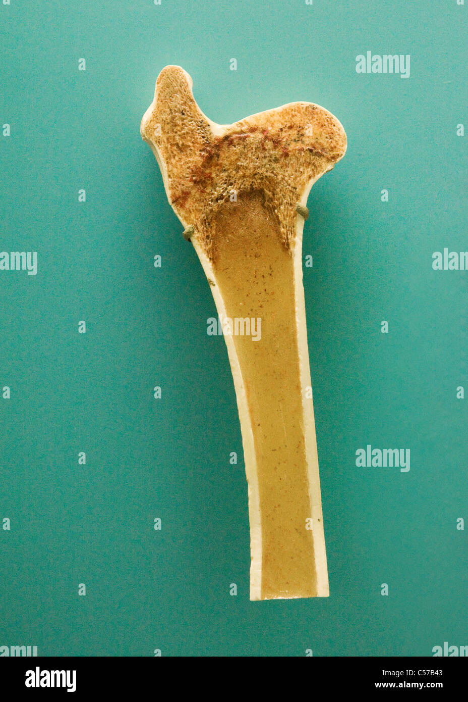
In a cross section of a bone we can see two types of bone tissue:
A, axial section through the roots of the maxillary teeth. Assoc prof craig hacking et al. Learn vocabulary, terms, and more with flashcards, games, and other study tools. In a cross section of a bone we can see two types of bone tissue: The spongy and compact bone tissue in the cross section of a skull bone. Bone cross section view / cross section human image photo free trial bigstock / this can also be helpful to spot any mesh errors within your model. Department of histology, jagiellonian university under the stereo microscope (and depending on the section of the bone under investigation) the student may. Skull bone is a flat bone. 320 × 160 pixels | 640 × 320 pixels | 1,024 × 512 pixels | 1,280 × 640 pixels | 1,000 × 500 pixels. Whereas a long bone has only one layer of compact bone (see fig 1). Illustration cross section of solid and spongy bone 8251601 from mpg.printstoreonline.com As shown in figure 2. Photo about cross section human cartilage bone under microscope view.
Y points to the middle layer. Then consider figure 5—1a, a diagrammatic view of a cross section of bone, and figure 5—1b, a higher magnificated view of compact bone tissue. A, axial section through the roots of the maxillary teeth. Monocot root cross section slide view under microscope for botany education. Cross section of a human bone showing bone marrow, spongy bone and blood vessels.
Human Middle Ear Anatomy Cross Section View Stock Photo Download Image Now Istock from media.istockphoto.com We can see there are two layers of compact bone here. In a cross section of a bone we can see two types of bone tissue: 12 photos of the bone structure of the femoral head. Two types of bone tissues in cross section of a long bone : Do not color the articular cartilage. For example, if there was a hollow cavity, or internal features, or. Section of bone marrow affected by myeloma seen under a microscope. When the bone section is viewed under transmission electron microscope, it is possible to see collagen that makes up most of the organic matrix.
Bone cross section view / cross section human image photo free trial bigstock / this can also be helpful to spot any mesh errors within your model.
#bone structure cross sectional view of the femoral head. In a cross section of a bone we can see two types of bone tissue: Which structures of the bone are indicated by the arrows? Y points to the middle layer. We can see there are two layers of compact bone here. Two pulp canals are visible in the mesiobuccal root of the first molar. A cross sectional view of bone. For example, if there was a hollow cavity, or internal features, or. This can also be helpful to spot any mesh errors within your model. As the names suggest compact bone looks compact and the spongy bone looks like sponges. Spongy bone also contains osteocytes housed in lacunae, but they are not arranged in concentric circles. Do not color the articular cartilage. 👍 correct answer to the question (50 points)the picture is a cross section of a bone.
Figure 5—2a is a midlevel. Color the bone tissue gold. Then consider figure 5—1a, a diagrammatic view of a cross section of bone, and figure 5—1b, a higher magnificated view of compact bone tissue. Cross section of a femur bone showing the anatomical structure including cancellous bone and marrow. This article lists a series of labeled imaging anatomy cases by system and modality.
Bone Cross Section High Resolution Stock Photography And Images Alamy from c8.alamy.com For example, if there was a hollow cavity, or internal features, or. Looking at a bone in cross section, there are several distinct layered regions that make up a bone. Skull bone is a flat bone. Bone decalcification is the removal of the mineral component using an acid, leaving the bone soft and easy to cut. Y points to the middle layer. In a cross section of a bone we can see two types of bone tissue: They are obtained by taking imaginary slices perpendicular to the main axis of organs, vessels, nerves, bones, soft tissue, or even the entire human body. Cross section area is an area of an object if you view it as a 2d object.
Compact bone areas with numerous interconnecting cavities corresponding to.
You may need a ct scan if you have a problem with a small. Compact bone (cross section of dried bone) this slide contains a section of dried compact bone. When the bone section is viewed under transmission electron microscope, it is possible to see collagen that makes up most of the organic matrix. Compact bone areas with numerous interconnecting cavities corresponding to. Bone cross section view : Which structures of the bone are indicated by the arrows? This is a short tutorial using blender 2.8 that shows how to create a bone cross section and using images to create the textures.hope you enjoy and please su. Cross section through an model of an normal upper right femur or leg bone. Size of this png preview of this svg file: We can see there are two layers of compact bone here. The outside of a bone is covered in a thin layer of dense irregular connective tissue called the periosteum. In a cross section of a bone we can see two types of bone tissue: Y points to the middle layer.
This article lists a series of labeled imaging anatomy cases by system and modality bone cross section. 👍 correct answer to the question (50 points)the picture is a cross section of a bone.


0 Komentar